Soubor:Tubal Pregnancy with embryo.jpg
Z Multimediaexpo.cz
(Rozdíly mezi verzemi)
(Fotografie + + pochází z Wikimedia Commons, kde má status – Kategorie:CC fotografie) |
(+ Doplnění) |
||
| Řádka 1: | Řádka 1: | ||
| - | Fotografie + | + | Fotografie + Description: Human Embryo (7th week of pregnancy, 5th week p.o.) |
| + | * This photo of an opened oviduct with an ectopic pregnancy features a spectacularly well preserved 10-millimeter embryo. It is uncommon to see any embryo at all in an ectopic, and for one to be this well preserved (and undisturbed by the prosector's knife) is quite unusual. | ||
| + | * Even an embryo this tiny shows very distinct anatomic features, including tail, limb buds, heart (which actually protrudes from the chest), eye cups, cornea/lens, brain, and prominent segmentation into somites. The gestational sac is surrounded by myriad chorionic villi resembling elongated party balloons. This embryo is about five weeks old (or seven weeks in the biologically misleading but eminently practical dating system used in obstetrics). | ||
| + | * The photo was taken on Kodak Elite 200 slide film, with a Minolta X-370 camera and 100mm f/4 Rokkor bellows lens at near-full extension. The formalin-fixed specimen was immersed in tap-water and pinned to a tray lined with black velvet. The exposure was 1/4 second at f/8. | ||
| + | * Date: First published: October 12, 2001 | ||
| + | * Source: (2014). "Tubal pregnancy with embryo". WikiJournal of Medicine 1 (2): 7. DOI:10.15347/wjm/2014.007. | ||
| + | * Author: Ed Uthman, MD (Flickr, Wikipedia) | ||
| - | + pochází z Wikimedia Commons, kde má status – | + | + pochází z Wikimedia Commons, kde má status – This work has been released into the public domain by its author, Euthman at English Wikipedia. |
| - | [[Kategorie: | + | [[Kategorie:PD fotografie]] |
Aktuální verze z 25. 10. 2017, 15:52
Fotografie + Description: Human Embryo (7th week of pregnancy, 5th week p.o.)
- This photo of an opened oviduct with an ectopic pregnancy features a spectacularly well preserved 10-millimeter embryo. It is uncommon to see any embryo at all in an ectopic, and for one to be this well preserved (and undisturbed by the prosector's knife) is quite unusual.
- Even an embryo this tiny shows very distinct anatomic features, including tail, limb buds, heart (which actually protrudes from the chest), eye cups, cornea/lens, brain, and prominent segmentation into somites. The gestational sac is surrounded by myriad chorionic villi resembling elongated party balloons. This embryo is about five weeks old (or seven weeks in the biologically misleading but eminently practical dating system used in obstetrics).
- The photo was taken on Kodak Elite 200 slide film, with a Minolta X-370 camera and 100mm f/4 Rokkor bellows lens at near-full extension. The formalin-fixed specimen was immersed in tap-water and pinned to a tray lined with black velvet. The exposure was 1/4 second at f/8.
- Date: First published: October 12, 2001
- Source: (2014). "Tubal pregnancy with embryo". WikiJournal of Medicine 1 (2): 7. DOI:10.15347/wjm/2014.007.
- Author: Ed Uthman, MD (Flickr, Wikipedia)
+ pochází z Wikimedia Commons, kde má status – This work has been released into the public domain by its author, Euthman at English Wikipedia.
Historie souboru
Kliknutím na datum a čas se zobrazí tehdejší verze souboru.
| Datum a čas | Náhled | Rozměry | Uživatel | Komentář | |
|---|---|---|---|---|---|
| současná | 25. 10. 2017, 15:18 | 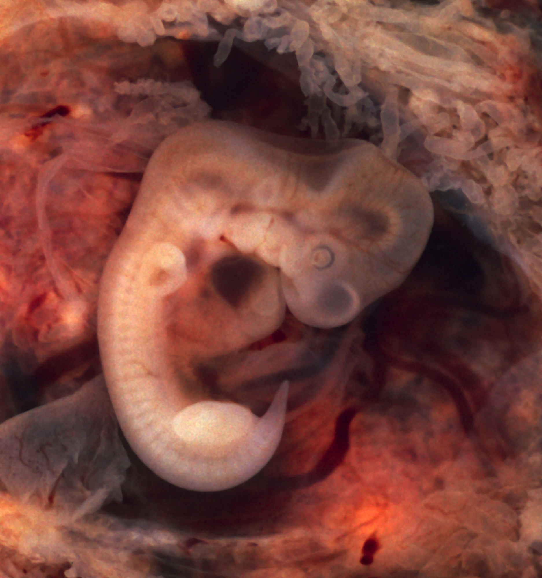 | 1 874×2 000 (1,48 MB) | Sysop (diskuse | příspěvky) | (Fotografie + + pochází z Wikimedia Commons, kde má status – Kategorie:CC fotografie ) |
- Editovat tento soubor v externím programu (Více informací najdete v nápovědě pro nastavení.)
Odkazy na soubor
Na soubor odkazuje tato stránka:


